
Immunoassays are indispensable tools in immunology research, using highly specific antibody–target interactions to detect and measure analytes in complex samples. These assays use immune-based reagents to produce a detectable signal, helping scientists study biological targets, monitor immune signalling pathways, and profile cells. As a result, immunoassays are widely applied to track immune responses to pathogens, study autoimmune disease progression, evaluate vaccine efficacy, and support post-transplant monitoring.
Among immunoassays, the enzyme-linked immunosorbent assay (ELISA) is perhaps the best known. It’s commonly used in routine laboratory workflows to detect and quantify protein-based analytes such as cytokines, antibodies, and antigens. However, despite its versatility, ELISA is not always the best fit for every immunology question, particularly when the biology involves rare events or subtle functional responses.
Immunology is a nuanced field where low-frequency responder cells and complex maturation patterns can be difficult to capture using bulk measurements alone. In the right context, the enzyme-linked immunospot (ELISpot) assay can provide greater resolution and a more functionally informative view of immune activity by measuring secretion at the single-cell level. This article outlines the key similarities and differences between ELISA and ELISpot and offers guidance to help researchers choose the most appropriate assay for their application.
An ELISA is a plate-based immunoassay used to detect and quantify specific proteins in liquid samples such as serum, plasma, or cell culture supernatants. ELISAs translate antibody–target binding into a measurable signal using an enzyme “reporter” that generates a colour change or a chemiluminescent/fluorescent readout. In most ELISA formats, the strength of the signal is proportional to the amount of target present, allowing researchers to compare samples and, when used with standards, calculate concentrations.1
Most ELISA workflows follow the same core steps: (1) immobilise a binding partner on the surface of a microplate well, (2) add the sample so the target can bind, (3) wash away unbound material, (4) add a detection system to label what’s been captured, and (5) add a substrate to produce a measurable signal.2
The step where a detection antibody is introduced is essential. The detection antibody binds specifically to the captured target and “labels” it so the assay can be visualised and quantified. In many setups, this detection antibody carries an enzyme itself, or it is recognised by an enzyme-linked secondary antibody. After excess antibody is washed away, a substrate is added; the enzyme converts this substrate into a coloured (or light-emitting) product that can be read on a microplate reader. This approach enhances sensitivity through enzymatic signal amplification. In sandwich formats, dual-antibody recognition of distinct epitopes adds an additional layer of selectivity.
The workflow explained above most closely describes a sandwich-style ELISA, where the target is captured by an immobilised antibody and then detected by a second antibody binding to a different epitope on the same target.2 However, not all ELISA assays are designed this way—different formats are used depending on whether you’re detecting an antigen or an antibody, the complexity of your sample, and the sensitivity you need.
Direct ELISA
The antigen is immobilised on the plate and detected with an enzyme-conjugated primary antibody.3 This format is fast with fewer steps, but can be less sensitive because there is no secondary amplification.

Indirect ELISA
The antigen is immobilised on the plate, an unlabelled primary antibody binds the antigen, and an enzyme-linked secondary antibody binds the primary antibody.4 This is commonly used for detecting antibodies in serum and often increases sensitivity through signal amplification.

Sandwich ELISA
A capture antibody is immobilised on the plate, the target antigen from the sample is captured, and a second (detection) antibody binds the target.5 This format is highly specific and well-suited to complex samples because the target is “sandwiched” between two antibodies.

Competitive ELISA
Sample antigen competes with a labelled antigen (or competes for antibody binding).6 Signal intensity is inversely proportional to the amount of target in the sample. This format is useful for small molecules or targets with only one available antibody-binding site.

ELISpot is an enzyme-linked immunoassay designed to detect and enumerate individual cells that are actively secreting a target protein, rather than measuring total analyte concentration.7 Although ELISpot and ELISA share core antibody-based detection principles, ELISpot protocols and use cases are distinct because the readout is a single-cell secretion event captured in place.
ELISpot is typically performed in 96-well plates or 8-well strips that contain a specialised membrane, most commonly polyvinylidene difluoride (PVDF) or nitrocellulose. This membrane captures secreted molecules close to the secreting cell, enabling highly sensitive, sandwich-based detection (Figure 1).8 Researchers can choose between pre-coated plates and manual coating with capture antibodies depending on their workflow. Pre-coated ELISpot plates arrive ready to use, which can be especially helpful for long-running studies or when sample timing is unpredictable. Alternatively, plates can be coated manually for full control over plate choice and assay setup.
In a standard ELISpot workflow, a defined number of viable cells is added to each well and then incubated to allow secretion of the analyte of interest.7,8 Secreted molecules are captured immediately by the immobilised antibodies directly beneath each cell, creating localised depositions at the point of secretion.
After incubation, cells are washed away while the captured analyte remains bound to the membrane. Detection antibodies are added to form a sandwich complex, followed by an enzyme-linked reporter and a chromogenic substrate (e.g., HRP- or ALP-based systems). The enzymatic reaction produces distinct coloured spots—each spot corresponding to one analyte-secreting cell—which can be counted manually or quantified using dedicated ELISpot readers.
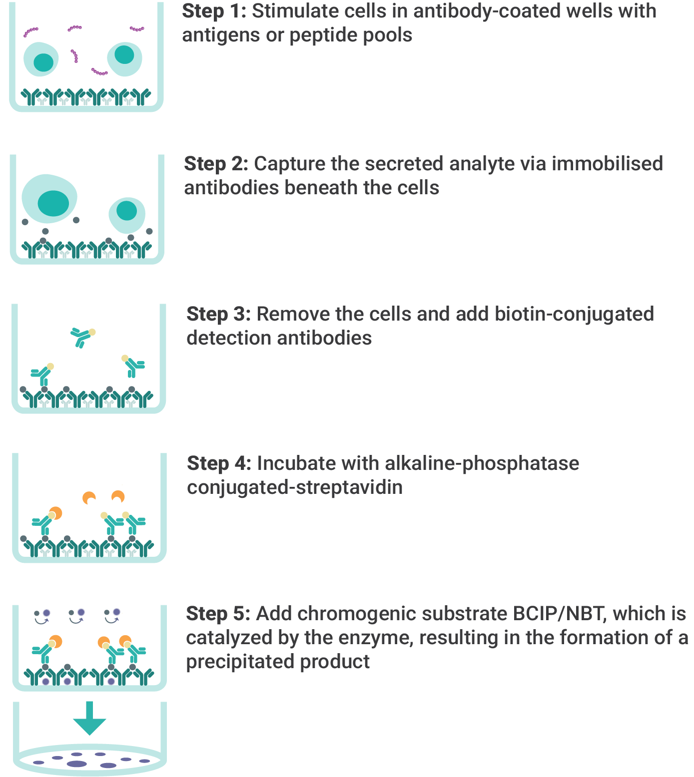
Figure 1. ELISpot assay steps.
At first glance, ELISA and ELISpot can look very similar: both are antibody-based immunoassays performed in microplate wells and rely on immobilised reagents and enzyme-driven signal generation. However, the two methods are designed to answer fundamentally different biological questions.
ELISA quantifies the total concentration of a target analyte in a liquid sample, such as serum, plasma, or cell culture supernatant, making it well-suited for measuring overall protein abundance across conditions or time points. The analyte concentration can reflect what cells have produced in the body or released into culture over time.
In contrast, ELISpot measures immune function at the single-cell level by counting how many individual cells are actively secreting a target molecule during an incubation step in the plate. ELISpot can detect a single cell secreting the target molecule amongst approximately 10⁶ cells, boasting a sensitivity that is 2-3 orders of magnitude higher than conventional ELISA. This is especially valuable for studying T cell behaviour and cytokine expression, including low-frequency responses that may be masked in bulk measurements.
To make the distinction clear, the key characteristics of ELISA and ELISpot are summarised below (Table 1).

Table 1. Comparison between ELISpot and ELISA.
ELISA is the better choice when your main goal is to measure the total amount of a target protein in a liquid sample. It’s designed to quantify analyte concentration (e.g., pg/mL or ng/mL) and is often the most direct option when you want a scalable, concentration-based readout across many samples.
ELISA is particularly appropriate in the following situations:
1) You need an absolute concentration readout
If your endpoint is “how much protein is present?” ELISA gives a quantitative concentration using standards and a standard curve, making it ideal for comparing levels across conditions and timepoints.
2) You’re measuring analytes in biofluids
When you want to assess what’s circulating in the body, such as cytokines, antibodies, or antigens in serum or plasma, ELISA is a straightforward way to quantify the in vivo output.
3) You’re running stimulated cell cultures and want the bulk response
ELISA is widely used to compare cytokine levels in supernatants after different stimulation conditions. It captures the overall amount produced by the culture over time, which is useful when “total output” is the key comparison.
4) You have limited access to viable cells
Because ELISA works with liquid samples (including stored serum/plasma or archived supernatants), it’s often more practical when live-cell assays aren’t possible or when samples have already been collected.
ELISpot assays offer single-cell resolution and sensitivity to rare cells. These assays are especially valuable when immune responses are rare or subtle, as they capture the secretion of proteins at the point of release and report results as discrete spot-forming units (SFU). This provides precise insights at the level of individual cells.
ELISpot is particularly appropriate in the following situations:
1) You need to quantify antigen-specific responding cells (especially T cells)
If your primary endpoint is the frequency of cells secreting a specific cytokine (e.g., IFN-γ, IL-2, TNF-α) after antigen stimulation, ELISpot directly answers that question by counting secreting cells rather than measuring total secretion across the whole culture. This is ideal for vaccine studies, infection research, and immune monitoring workflows where the “responder frequency” is the most informative readout.
2) You expect low-frequency responses and need high sensitivity
ELISpot is well-suited to detecting rare responder populations that may be masked in bulk assays, making it valuable when responder frequencies are expected to be low or your cell sample is limited. In sensitivity-focused applications, ELISpot can detect secretion from very small numbers of responding cells in a large background population.
3) You want functional insight
ELISA measures the total concentration of proteins in a sample, reflecting overall immune activity but not linking responses to individual cells. In contrast, the ELISpot quantifies the number of individual cells actively secreting a specific cytokine during the assay. This approach provides a direct measure of responder frequency, allowing researchers to differentiate whether the observed response comes from a small number of highly active cells or a larger population of moderately active ones.
4) You are already working with viable cells for parallel analyses
ELISpot requires viable cells as the assay input (commonly PBMCs, splenocytes, or other primary immune cells) and is designed around short-term culture and stimulation. It’s a good fit when you’re already culturing cells for parallel analyses (e.g., flow cytometry phenotyping, activation marker panels, or cell sorting).
5) You want to use ELISpot alongside ELISA and flow cytometry
ELISpot can complement other immunoassays like ELISA and flow cytometry by addressing different aspects of the same question. ELISA shows how much protein is present overall in a sample after stimulation, while flow cytometry lets you quantify immune cell populations. ELISpot adds a complementary functional readout by counting how many cells are actively secreting the cytokine during the assay. Used together, these approaches help you connect magnitude, phenotype and subset attribution, and responder frequency.
Because ELISA and ELISpot answer different questions, their outputs look different, too. Understanding the typical readouts makes it easier to choose the right assay and interpret results with confidence.
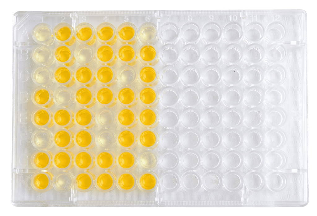
Typical ELISA readouts
ELISA results are usually reported as a concentration derived from a standard curve.
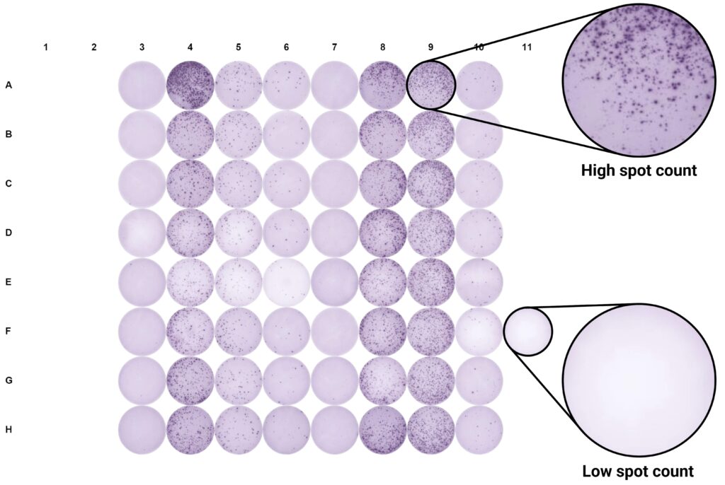
Typical ELISpot readouts
ELISpot results are reported as spot counts, where each spot represents an individual secreting cell event under the assay conditions.
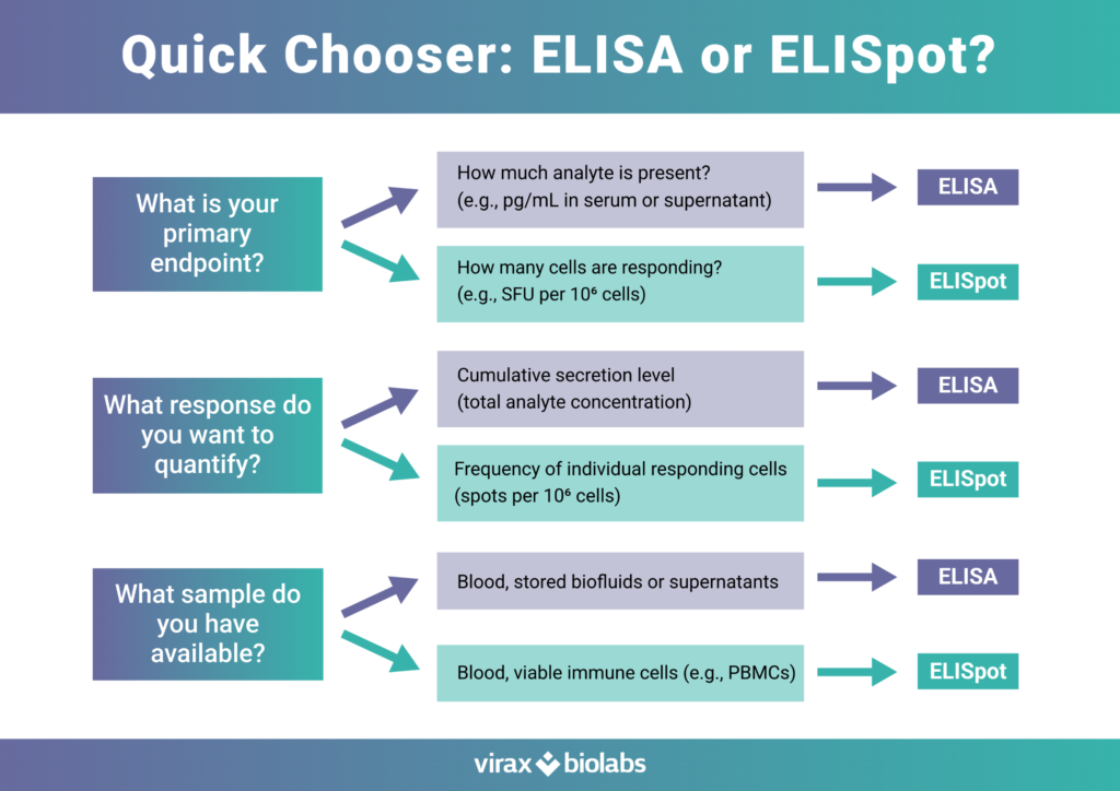
Choosing between ELISA and ELISpot comes down to what you need to learn from your experiment. If your goal is to quantify how much target analyte is present in a biofluid or supernatant, ELISA remains a robust and scalable option. If you need to understand how many cells are responding—especially when responses are rare, subtle, or antigen-specific—ELISpot offers single-cell functional resolution that bulk measurements can miss. Using the right assay for the right endpoint helps ensure your readout aligns with the biology you’re trying to capture and supports clearer, more confident decisions in immune monitoring, vaccine research, and translational studies.
Virax Biolabs offers a broad portfolio of ELISpot kits and services to support immune response profiling across diverse targets and study designs. Choose from coated and non-coated plate formats, and work with our team on custom assay design, using the latest peptide pool technology to achieve accurate T cell stimulation reflective of your research needs.
New to ELISpot?
We can help you get started without upfront infrastructure investment. Whether you need your samples analysed by our team or hands-on training at your site, we offer flexible support options for UK-based researchers.